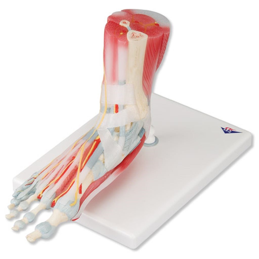
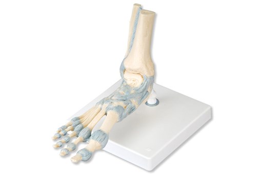
Foot Skeleton With Ligaments And Muscles
FRTH-1000360
$0
inc gst
Add to cart
In stock
Product overview
This 3B Scientific model is the best of its kind for quality and value.
This anatomically detailed model of the foot and lower leg can be disassembled into 6 removable parts for detailed study.
The model features not only the bones, but also the muscles, tendons, ligaments, nerves, arteries and veins.
The frontal view features the extensor muscles of the lower leg. The tendons can be followed on their passage under the transverse and crucial crural ligaments all the way to their insertion points. In addition, all tendon sheaths are visible.
On the dorsal portion of the model, the gastrocnemius muscle is removable to reveal deeper anatomical elements.
The sole of the foot is represented in three layers; the first layer displaying the flexor digitorum brevis. This muscle can be removed revealing the quadratus plantae, the tendon of the flexor digitorum longus, and the flexor hallucis muscle. This second layer is, in turn, removable to display even deeper anatomical details.
Size: 23 x 26 x 19cm. Weight: 1.1kg
You may also be interested in
inc gst
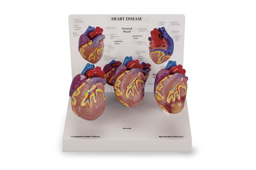
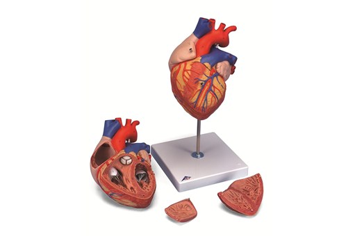
3 Piece Mini Heart Set Models
Specialist
Order
Order
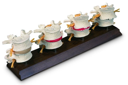
4th And 5th Lumbar Model
Specialist
Order
Order

Anthology of Anatomical Charts
Specialist
Order
Order
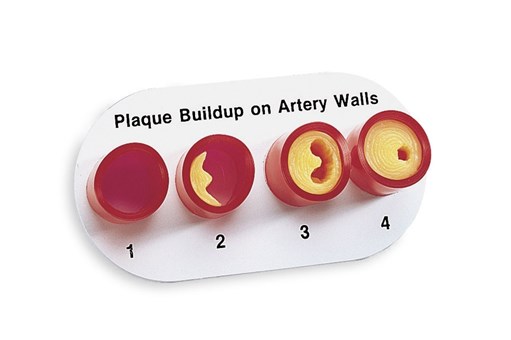
Artery Section Complete With Blockage
Specialist
Order
Order
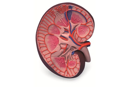
Basic Kidney Section Model
inc gst
Brain 2 Part
inc gst
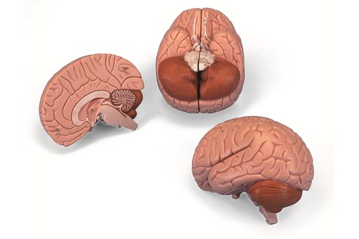
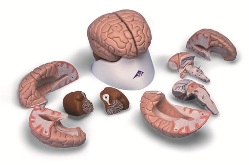
Brain 4 Part
inc gst
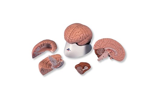
Brain Anatomy Model 8 Part
Specialist
Order
Order
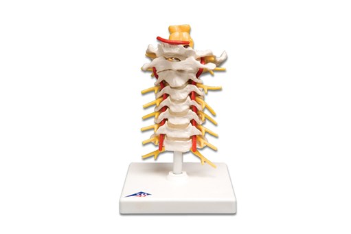
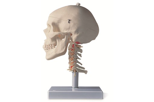
Cervical Spinal Column Model
inc gst
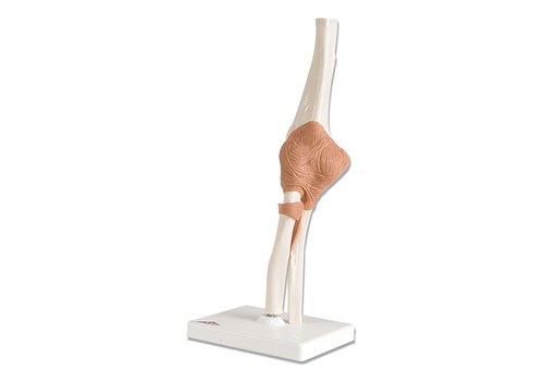
Classic Flexible Elbow Joint Model
inc gst
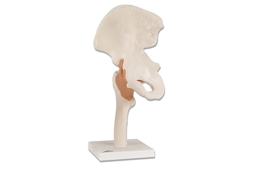
Classic Flexible Hip Joint Model
inc gst
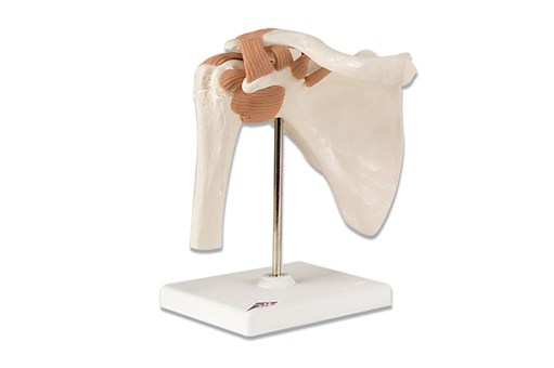
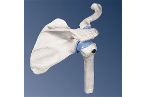
Classic Flexible Shoulder Joint Model
Specialist
Order
Order
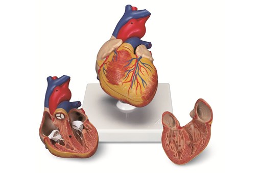
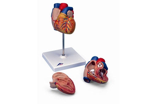
Classic Heart 2 Part
inc gst
Classic Heart with Conducting System 2 Part
inc gst
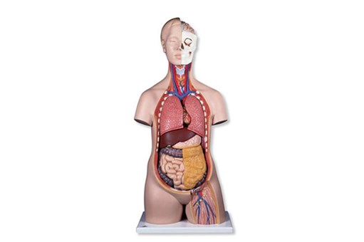
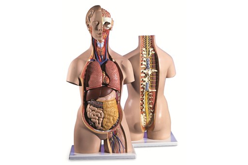
Classic Unisex Torso 12 Part
Specialist
Order
Order
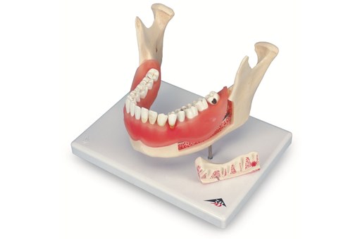
Clear Human Jaw With Teeth
Specialist
Order
Order
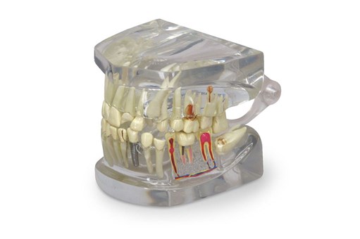
Dental Disease Model
Specialist
Order
Order
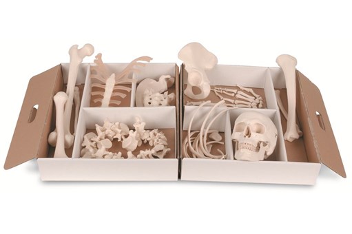
Disarticulated Half Skeleton Model
inc gst
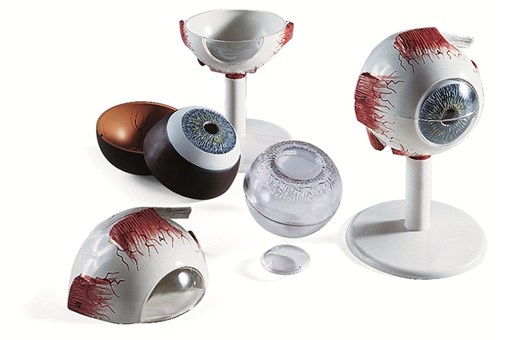
Eye Anatomy Model 3 Times Full Size 6 Part
inc gst
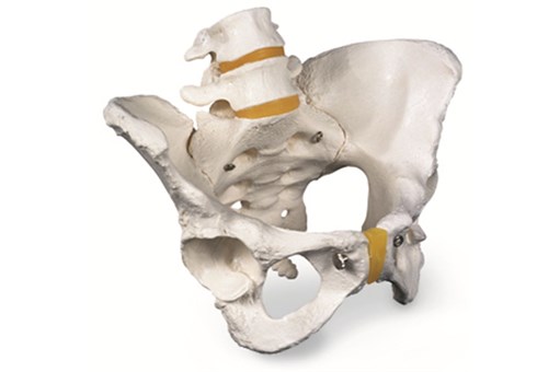
Female Pelvic Skeleton Model
inc gst
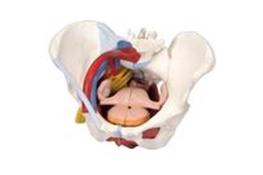
Female Pelvis 6 Part
Specialist
Order
Order
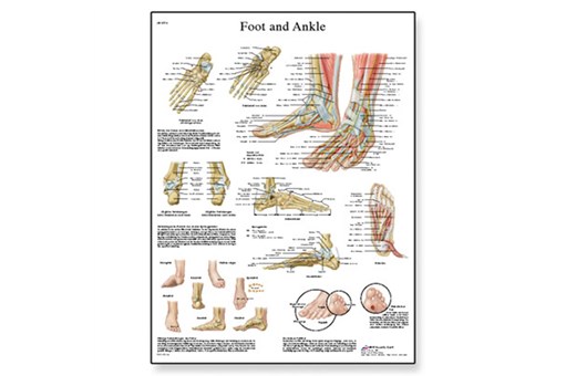
Foot And Joints Of The Foot Chart
inc gst
Foot Skeleton Model With Ligaments
inc gst
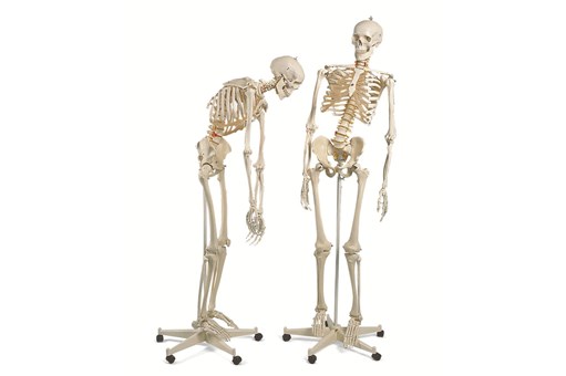
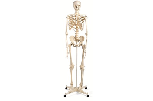
Fred Flexible Skeleton Model On 5ft Roller Stand
inc gst
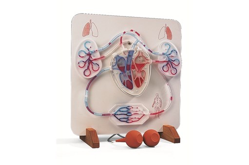
Functional Heart And Circulatory System Model
Specialist
Order
Order
Functional Larynx 2.5 Times Full Size
Specialist
Order
Order
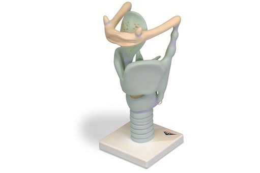
Functional Larynx 3 Times Life Size
Specialist
Order
Order
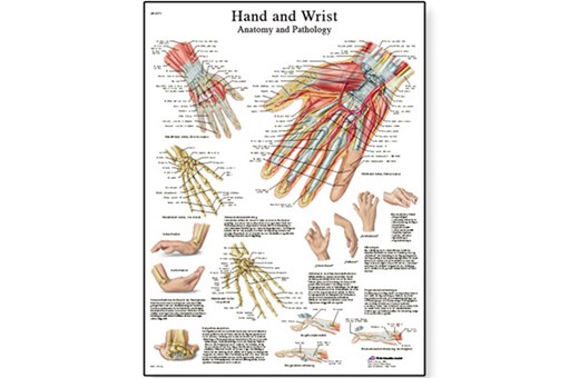
Hand And Wrist Chart
inc gst
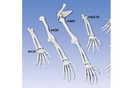
Hand Skeleton Ulna And Radius Flex
Specialist
Order
Order
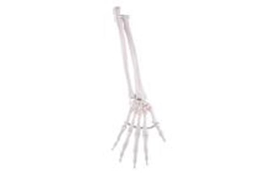
Hand Skeleton With Bone Names
Specialist
Order
Order
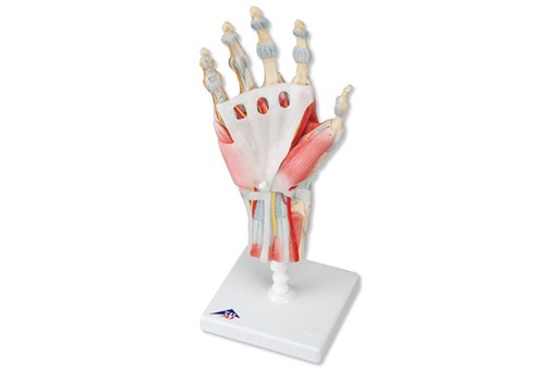
Hand Skeleton With Ligaments And Muscles
Specialist
Order
Order
Heart 2 Times Life Size 4 Part
Specialist
Order
Order
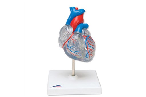
Heart Model
inc gst
Heart Model 2 Part
Specialist
Order
Order

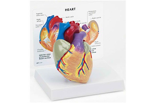
Heart With Oesophagus And Trachea
Specialist
Order
Order
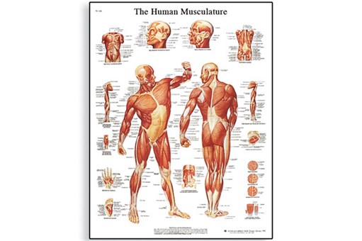
Human Musculature Chart
Specialist
Order
Order
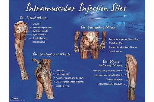
Intramuscular Injection Sites Poster
Specialist
Order
Order

Kidney Model With Pathologies
Specialist
Order
Order
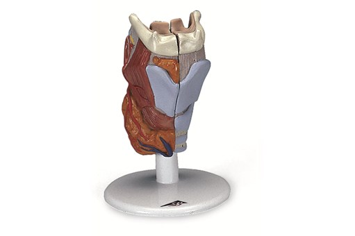
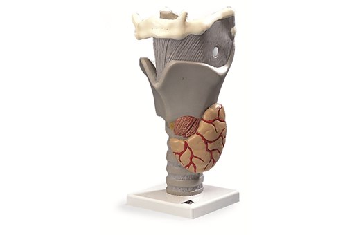
Larynx 2 Part
Specialist
Order
Order
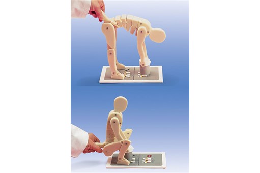
Lifting Demonstration Figure
Specialist
Order
Order
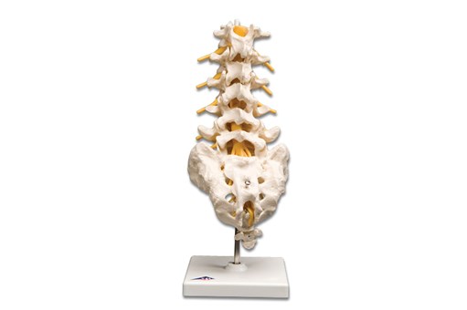
Lumbar Spinal Column Model
Specialist
Order
Order
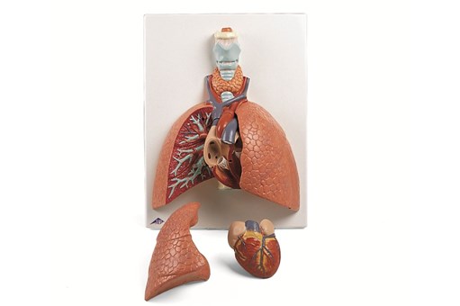
Lung Model With Larynx 5 Part
Specialist
Order
Order
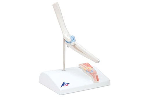
Mini Elbow Joint with Cross Section
inc gst
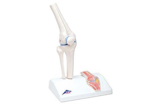
Mini Knee Joint with Cross Section
inc gst
Mini Shoulder Joint with Cross Section
inc gst
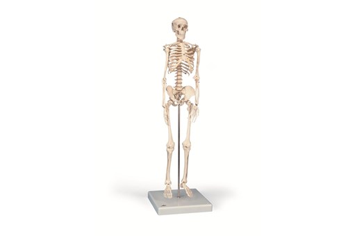
Mini Skeleton 'Shorty' Model
inc gst
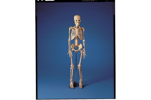
Mr Thrifty 84cm Skeleton Model
inc gst
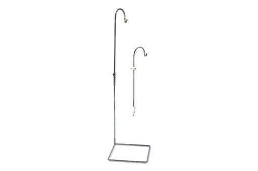
Multifunctional Stand
Specialist
Order
Order
Nasco Skin Cancer Model
Specialist
Order
Order
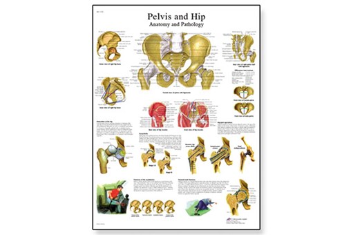
Pelvis And Hip Chart
Specialist
Order
Order
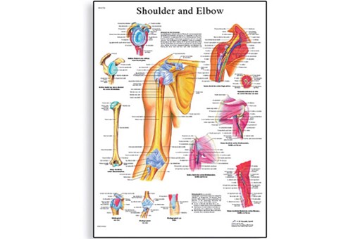
Shoulder And Elbow Chart
Specialist
Order
Order
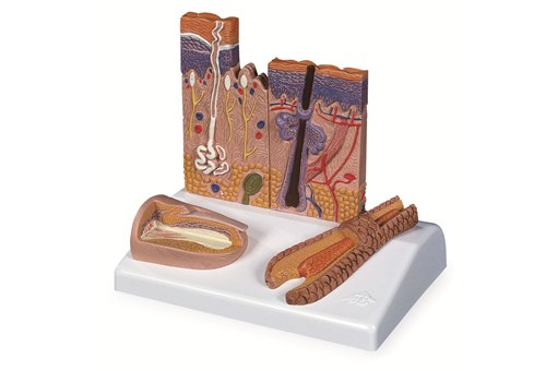
Skin Model - 3 Part
Specialist
Order
Order
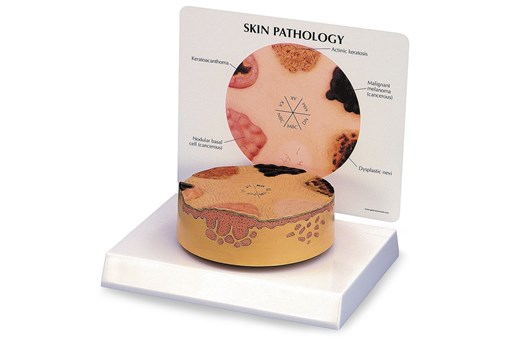
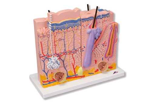
Skin Section Model
Specialist
Order
Order
Skull Model - 4 Part
Specialist
Order
Order
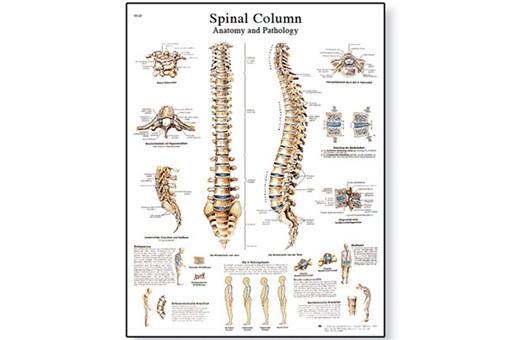
Spinal Column Chart
Specialist
Order
Order
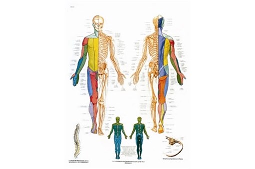
Spinal Nerves Chart
Specialist
Order
Order
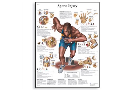
Sports Injuries Chart
Specialist
Order
Order
Stan Classical Skeleton Model On 5 ft Roller Stand
inc gst
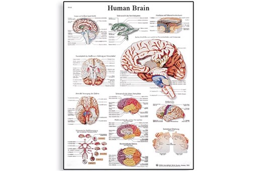
The Human Brain Chart
inc gst
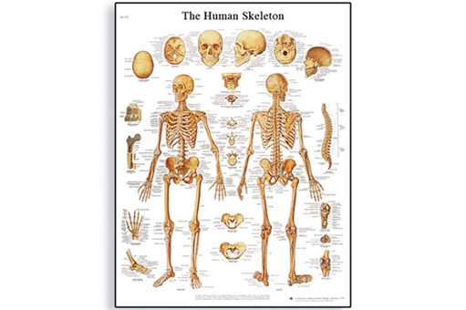
The Human Skeleton Chart
inc gst
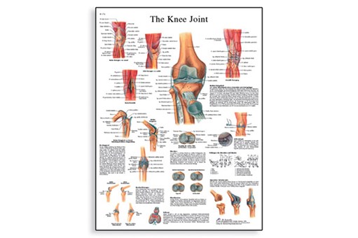
The Knee Joint Chart
Specialist
Order
Order
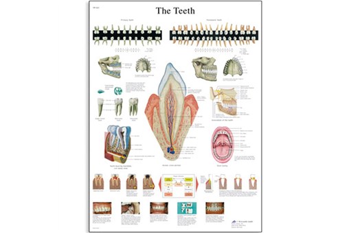
The Teeth Chart
Specialist
Order
Order
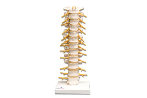
Thoracic Spinal Column Model
Specialist
Order
Order