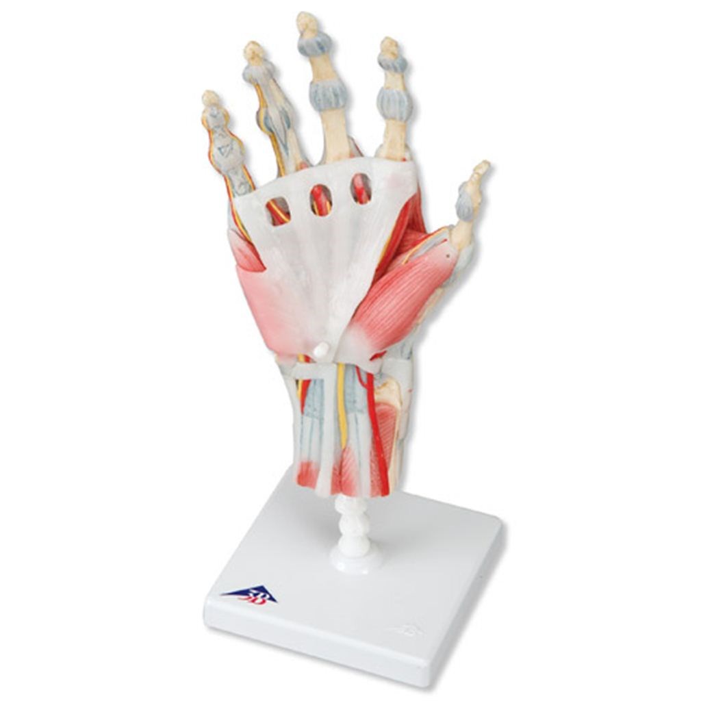
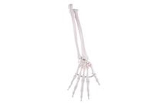
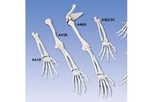
Hand Skeleton With Ligaments And Muscles
FRTH-1000358
Specialist Order
This product is available for special order only, call Customer Services on +64 9 969 2700 or Freephone 0508 414 564 to enquire or click Enquire Now to send us a message.
Product overview
The bones, muscles, tendons, ligaments, nerves, arteries and veins are all featured in this high quality 4 part model from 3B Scientific of the hand and lower forearm.
The dorsal side shows the extensor muscles as well as portions of the tendons at the wrist as they pass under the extensor retunaculum.
The palmar face of the hand is represented in three layers, the first two removable to allow detailed study of the deeper anatomical layer.
In addition, clinically important structures such as the median nerve and superficial palmar arterial arch can be examined in detail.
The deepest anatomical layer allows for study of the intrinsic muscles and deep palmar arterial arch in addition to other details.
Size: 33 x 12 x 12cm. Weight: 0.4kg
You may also be interested in
inc gst
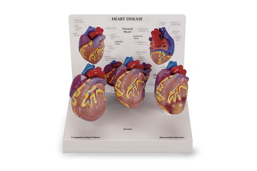
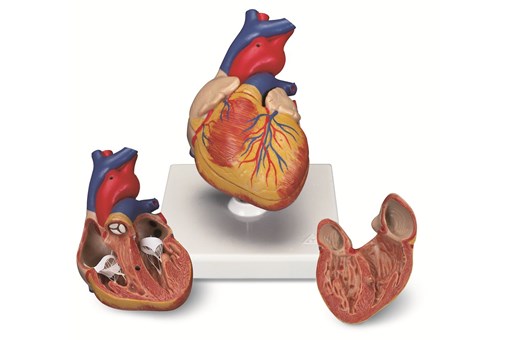
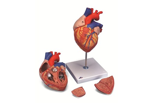
3 Piece Mini Heart Set Models
Specialist
Order
Order
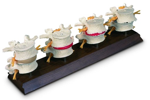
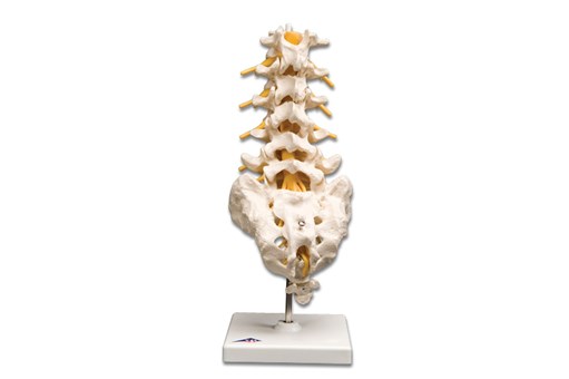
4th And 5th Lumbar Model
Specialist
Order
Order
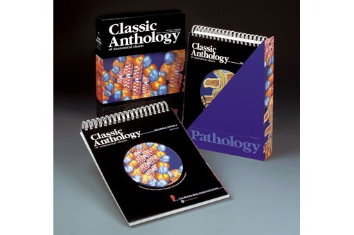
Anthology of Anatomical Charts
Specialist
Order
Order
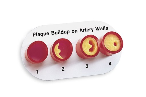
Artery Section Complete With Blockage
Specialist
Order
Order
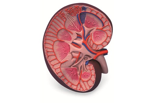
Basic Kidney Section Model
inc gst
Brain 2 Part
inc gst
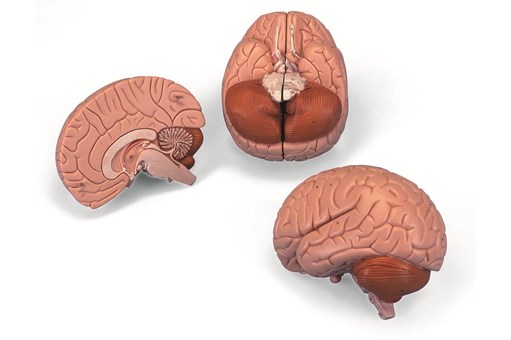
Brain 4 Part
inc gst
Brain Anatomy Model 8 Part
Specialist
Order
Order
Cervical Spinal Column Model
inc gst
Classic Flexible Elbow Joint Model
inc gst
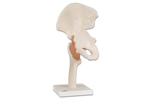
Classic Flexible Hip Joint Model
inc gst
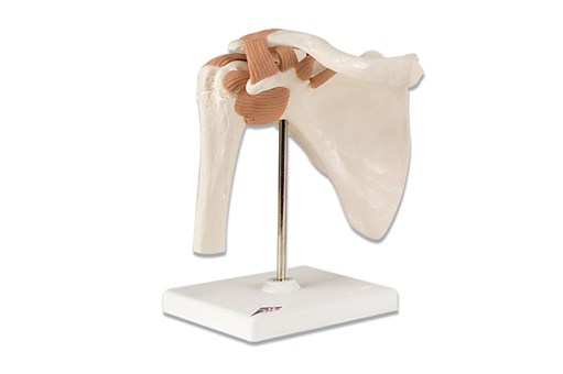
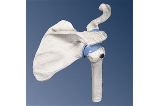
Classic Flexible Shoulder Joint Model
Specialist
Order
Order
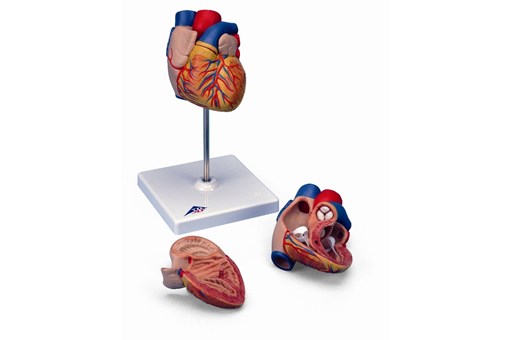
Classic Heart 2 Part
inc gst
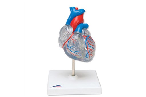
Classic Heart with Conducting System 2 Part
inc gst
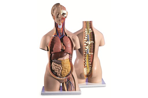
Classic Unisex Torso 12 Part
Specialist
Order
Order
Clear Human Jaw With Teeth
Specialist
Order
Order
Dental Disease Model
Specialist
Order
Order
Disarticulated Half Skeleton Model
inc gst
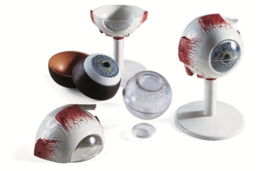
Eye Anatomy Model 3 Times Full Size 6 Part
inc gst
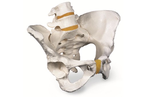
Female Pelvic Skeleton Model
inc gst
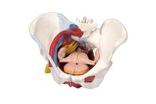
Female Pelvis 6 Part
Specialist
Order
Order
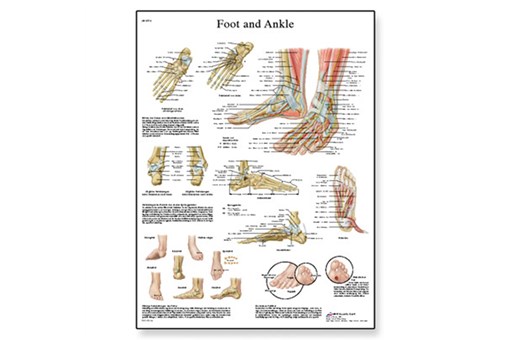
Foot And Joints Of The Foot Chart
inc gst
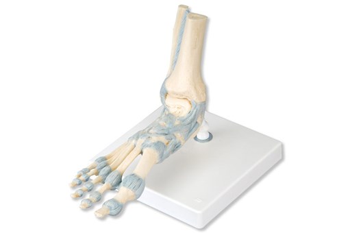
Foot Skeleton Model With Ligaments
inc gst
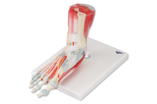
Foot Skeleton With Ligaments And Muscles
inc gst
Fred Flexible Skeleton Model On 5ft Roller Stand
inc gst
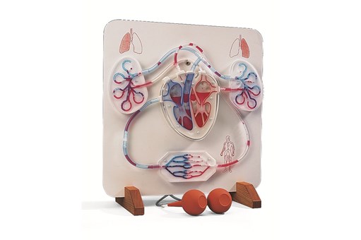
Functional Heart And Circulatory System Model
Specialist
Order
Order
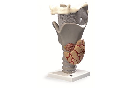
Functional Larynx 2.5 Times Full Size
Specialist
Order
Order
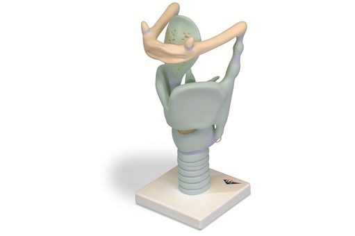
Functional Larynx 3 Times Life Size
Specialist
Order
Order
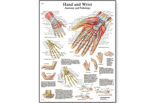
Hand And Wrist Chart
inc gst
Hand Skeleton Ulna And Radius Flex
Specialist
Order
Order
Hand Skeleton With Bone Names
Specialist
Order
Order
Heart 2 Times Life Size 4 Part
Specialist
Order
Order
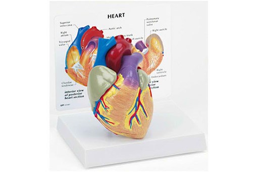
Heart Model
inc gst
Heart Model 2 Part
Specialist
Order
Order

Heart With Oesophagus And Trachea
Specialist
Order
Order
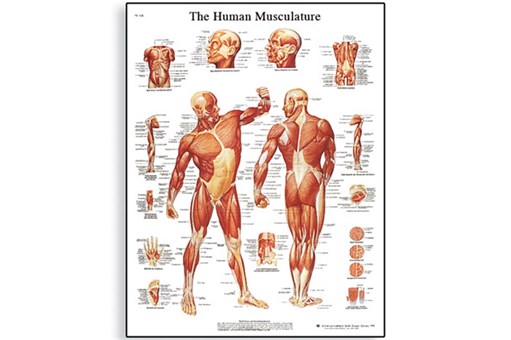
Human Musculature Chart
Specialist
Order
Order
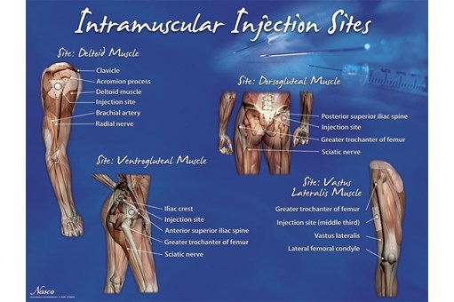
Intramuscular Injection Sites Poster
Specialist
Order
Order

Kidney Model With Pathologies
Specialist
Order
Order
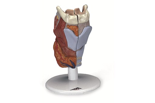
Larynx 2 Part
Specialist
Order
Order
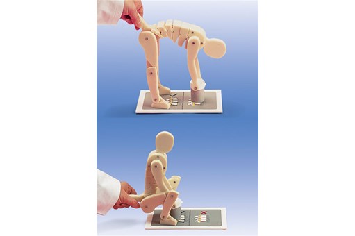
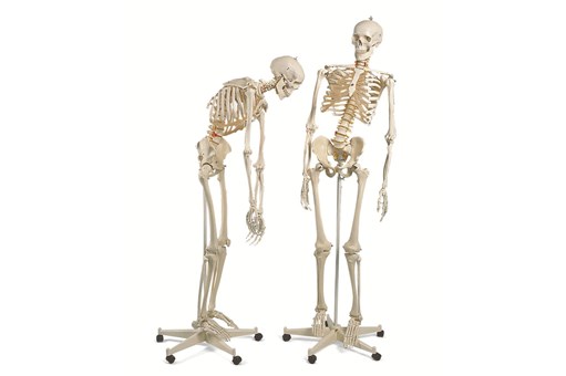
Lifting Demonstration Figure
Specialist
Order
Order
Lumbar Spinal Column Model
Specialist
Order
Order
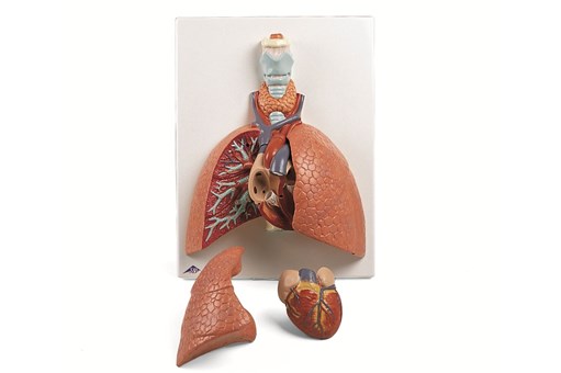
Lung Model With Larynx 5 Part
Specialist
Order
Order
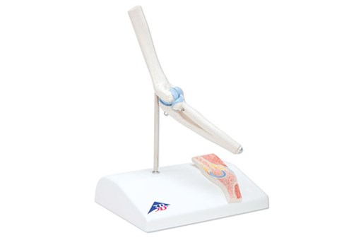
Mini Elbow Joint with Cross Section
inc gst
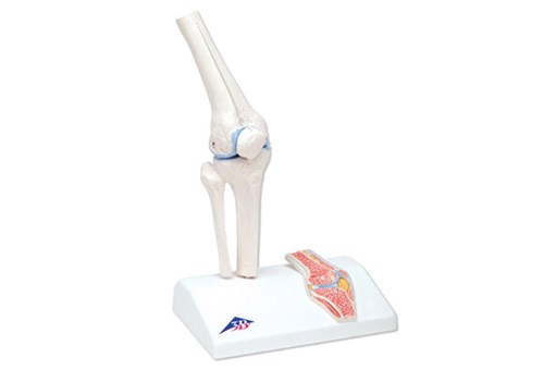
Mini Knee Joint with Cross Section
inc gst
Mini Shoulder Joint with Cross Section
inc gst
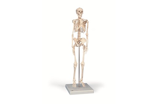
Mini Skeleton 'Shorty' Model
inc gst
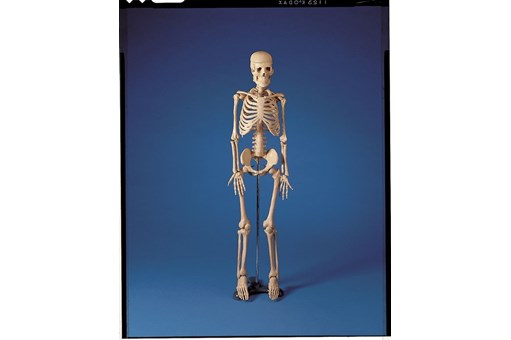
Mr Thrifty 84cm Skeleton Model
inc gst
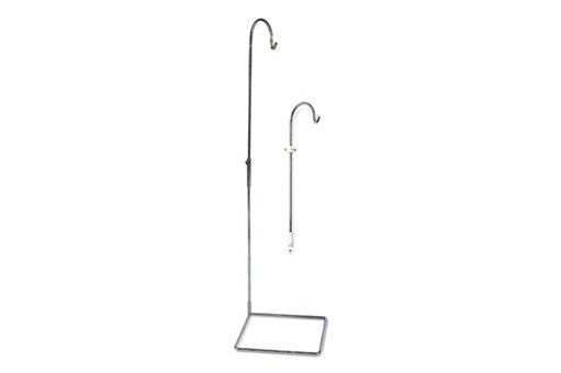
Multifunctional Stand
Specialist
Order
Order
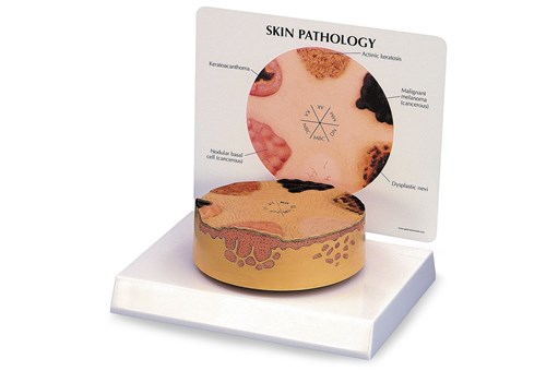
Nasco Skin Cancer Model
Specialist
Order
Order
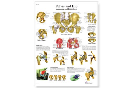
Pelvis And Hip Chart
Specialist
Order
Order
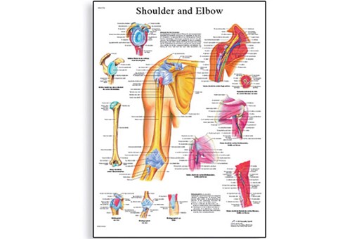
Shoulder And Elbow Chart
Specialist
Order
Order
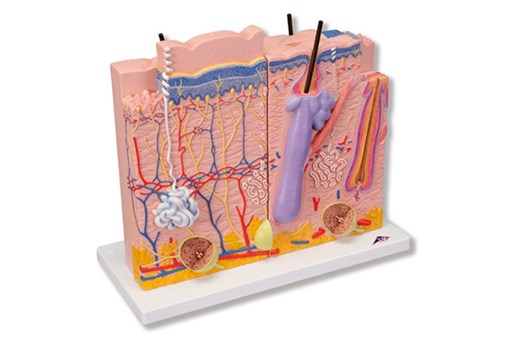
Skin Model - 3 Part
Specialist
Order
Order
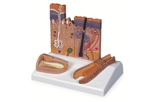
Skin Section Model
Specialist
Order
Order
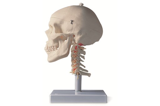
Skull Model - 4 Part
Specialist
Order
Order
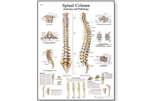
Spinal Column Chart
Specialist
Order
Order
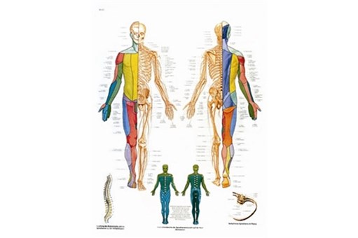
Spinal Nerves Chart
Specialist
Order
Order
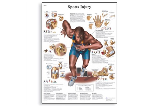
Sports Injuries Chart
Specialist
Order
Order
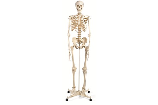
Stan Classical Skeleton Model On 5 ft Roller Stand
inc gst
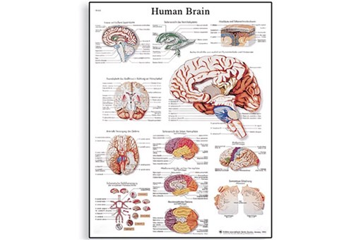
The Human Brain Chart
inc gst
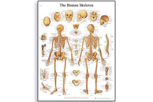
The Human Skeleton Chart
inc gst
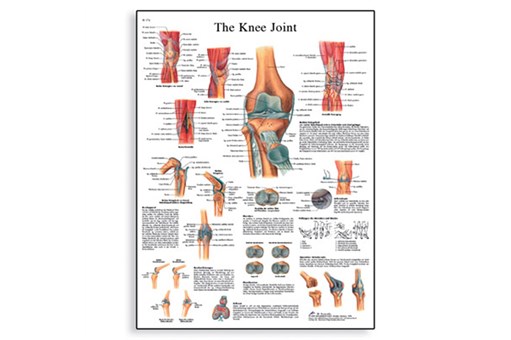
The Knee Joint Chart
Specialist
Order
Order
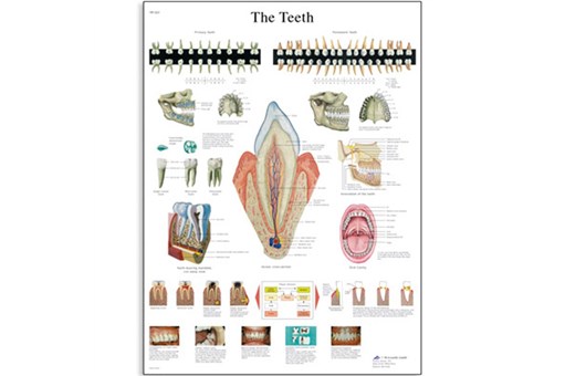
The Teeth Chart
Specialist
Order
Order
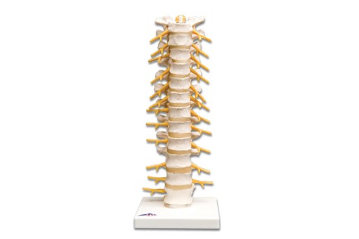
Thoracic Spinal Column Model
Specialist
Order
Order