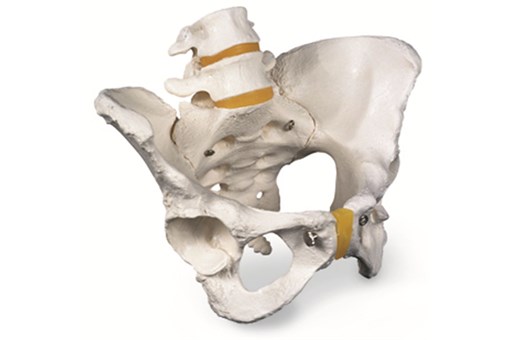
Anatomical / Skeletons
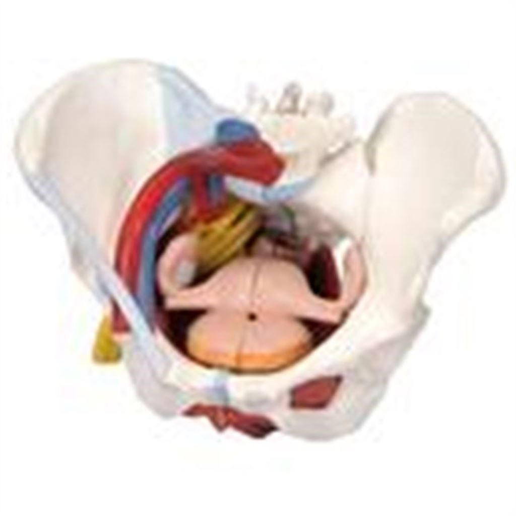
Female Pelvis 6 Part
FRTH-1000288
$0
inc gst
Add to cart
In stock
Product overview
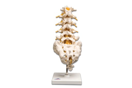
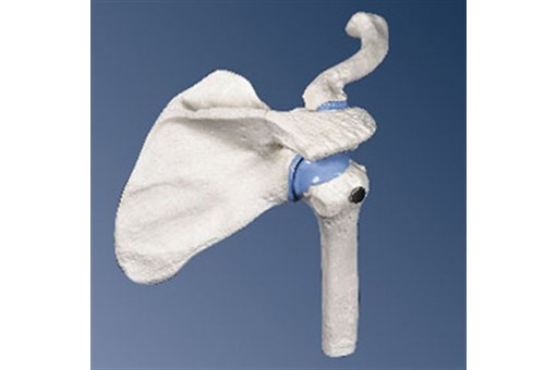
This 6 part model of a female pelvis represents detailed information about the topography of bones, ligaments, vessels, nerves, pelvic floor muscles and female pelvic organs.
It presents the whole pelvic floor with partially removable midsagitally sectioned external anal sphincter, external urethral sphincter, deep and superficial transverse perineal and bulbospongiosus.
Rectum, uterus with fallopian tubes, ovaries and vagina are also removable and can be disassembled into 2 halves by midsagital section.
The right pelvic half demonstrates the divisions and topographical anatomy of the common iliac artery, the external and internal artery and also of the common iliac vein and the external iliac vein.
The right sacral plexus, right sciatic nerve and right pudendal nerve are also shown.
Bones and ligaments presented are:
- 2 hip bones
- the pubic symphysis
- the sacrum and the coccyx
- the 5th lumbar vertebra with intervertebral disc
A midsagital section through the fifth lumbar vertebra, sacrum and coccyx allow both halves of the pelvis to be disassembled revealing a part of the cauda equina in the vertebral canal. The left half of the fifth lumbar vertebral body is removable.
The right half of this female pelvis shows the following pelvic ligaments
- the sacrotuberous ligament
- the inguinal ligament
- the sacrospinous ligament
- the anterior sacroiliac ligaments
- the iliolumbar ligament
- the anterior longitudinal ligament
- the interosseous sacroiliac ligament
- the posterior sacroiliac ligament
- the obturator
Dimensions: 19cm x 27cm x 19cm
Weight: 1.6kg
You may also be interested in
inc gst
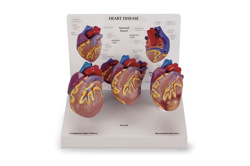
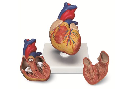
3 Piece Mini Heart Set Models
Specialist
Order
Order
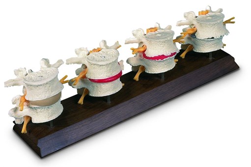
4th And 5th Lumbar Model
Specialist
Order
Order
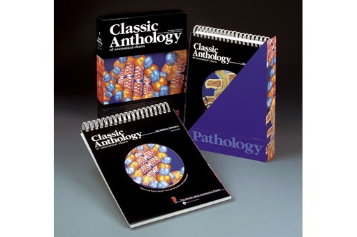
Anthology of Anatomical Charts
Specialist
Order
Order
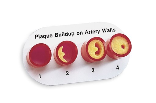
Artery Section Complete With Blockage
Specialist
Order
Order
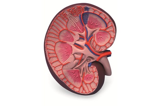
Basic Kidney Section Model
inc gst
Brain 2 Part
inc gst
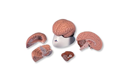
Brain 4 Part
inc gst
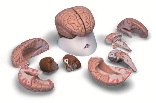
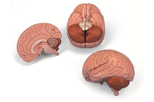
Brain Anatomy Model 8 Part
Specialist
Order
Order
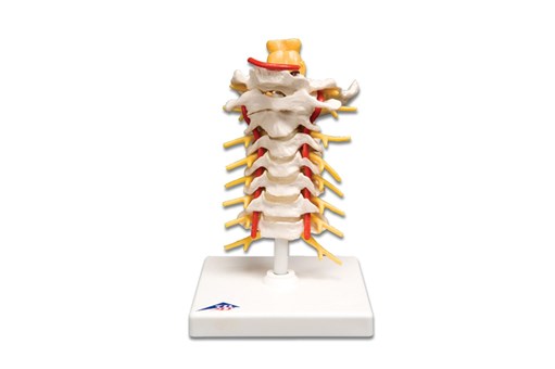
Cervical Spinal Column Model
inc gst
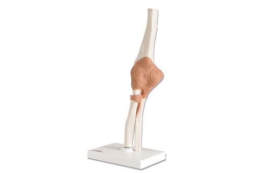
Classic Flexible Elbow Joint Model
inc gst
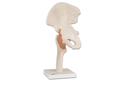
Classic Flexible Hip Joint Model
inc gst
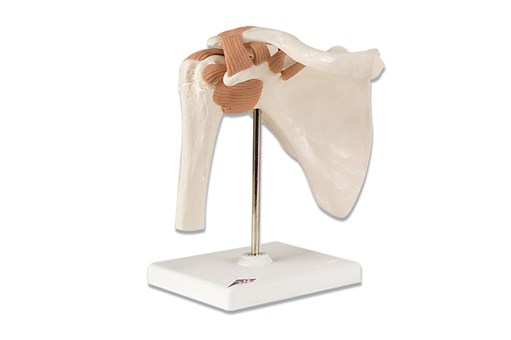
Classic Flexible Shoulder Joint Model
Specialist
Order
Order
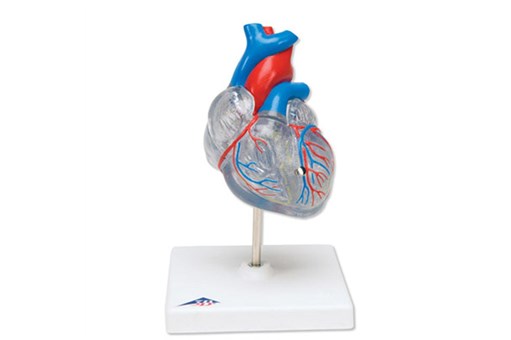
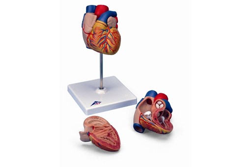
Classic Heart 2 Part
inc gst
Classic Heart with Conducting System 2 Part
inc gst
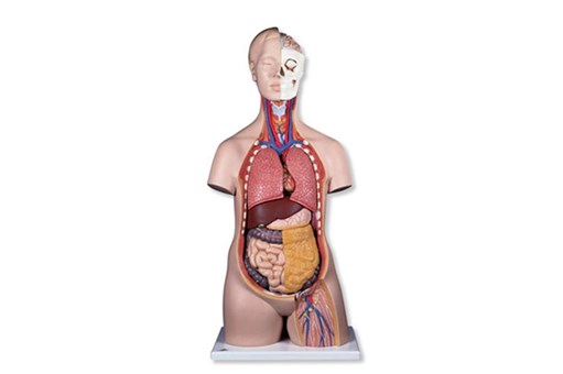
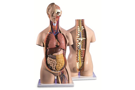
Classic Unisex Torso 12 Part
Specialist
Order
Order
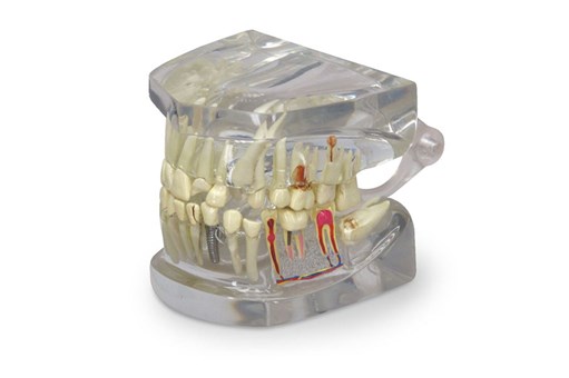
Clear Human Jaw With Teeth
Specialist
Order
Order
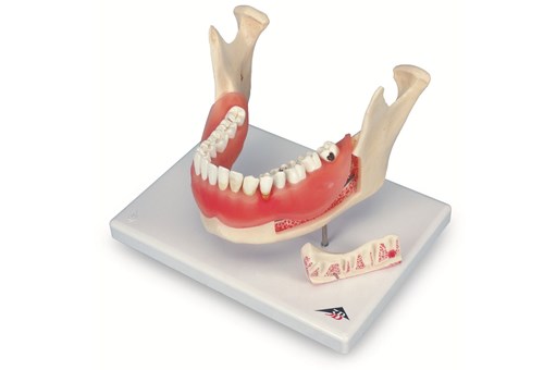
Dental Disease Model
Specialist
Order
Order
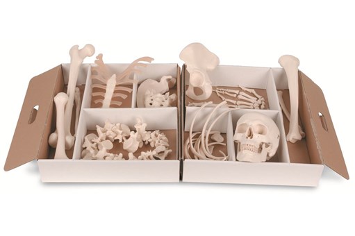
Disarticulated Half Skeleton Model
inc gst
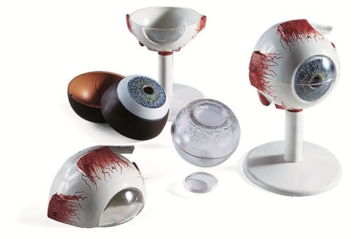
Eye Anatomy Model 3 Times Full Size 6 Part
inc gst
Female Pelvic Skeleton Model
Specialist
Order
Order
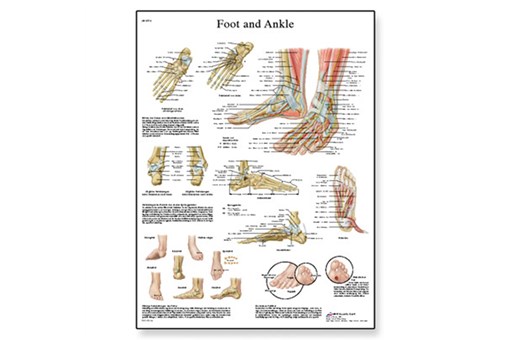
Foot And Joints Of The Foot Chart
inc gst
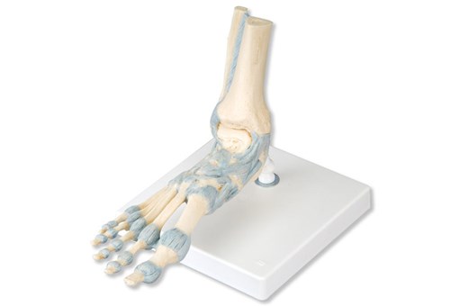
Foot Skeleton Model With Ligaments
inc gst
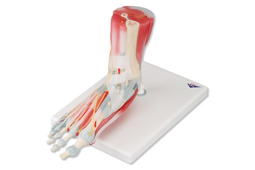
Foot Skeleton With Ligaments And Muscles
inc gst
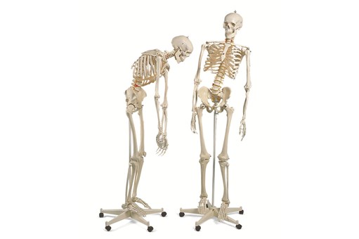
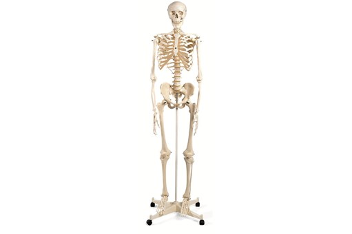
Fred Flexible Skeleton Model On 5ft Roller Stand
inc gst
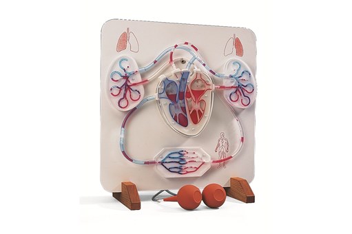
Functional Heart And Circulatory System Model
Specialist
Order
Order
Functional Larynx 2.5 Times Full Size
Specialist
Order
Order
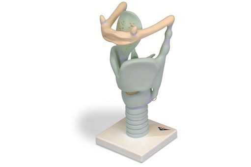
Functional Larynx 3 Times Life Size
Specialist
Order
Order
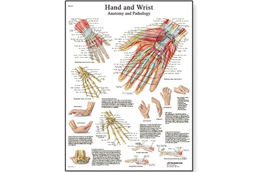
Hand And Wrist Chart
inc gst
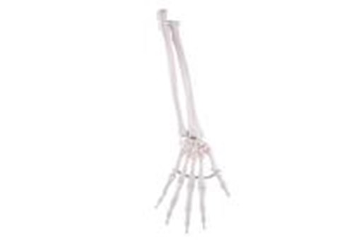
Hand Skeleton Ulna And Radius Flex
Specialist
Order
Order
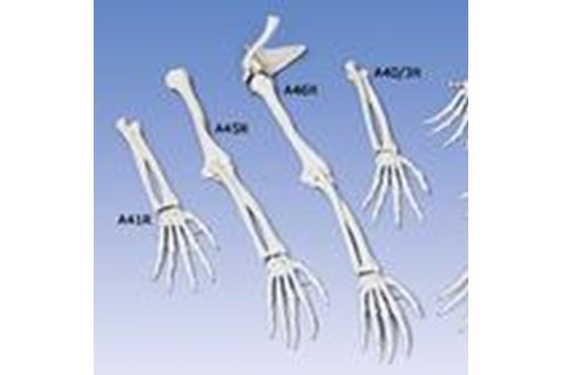
Hand Skeleton With Bone Names
Specialist
Order
Order
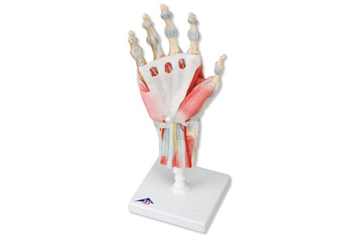
Hand Skeleton With Ligaments And Muscles
Specialist
Order
Order
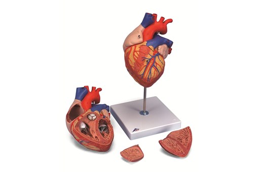
Heart 2 Times Life Size 4 Part
Specialist
Order
Order
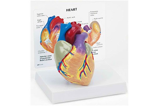
Heart Model
inc gst
Heart Model 2 Part
Specialist
Order
Order

Heart With Oesophagus And Trachea
Specialist
Order
Order
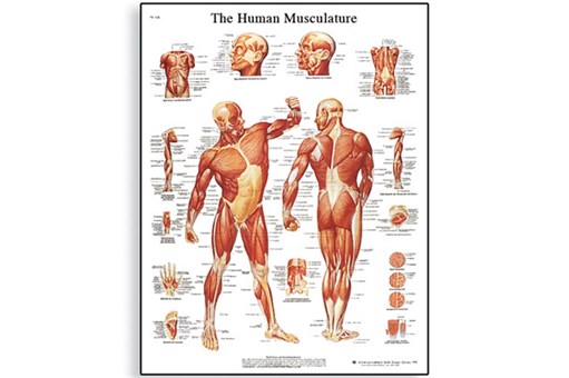
Human Musculature Chart
Specialist
Order
Order
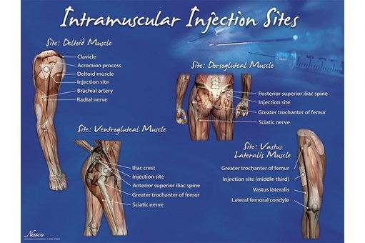
Intramuscular Injection Sites Poster
Specialist
Order
Order

Kidney Model With Pathologies
Specialist
Order
Order
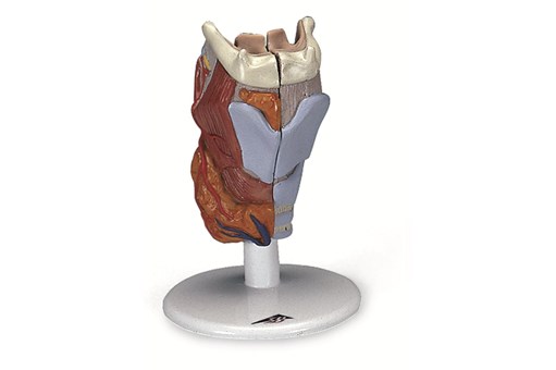
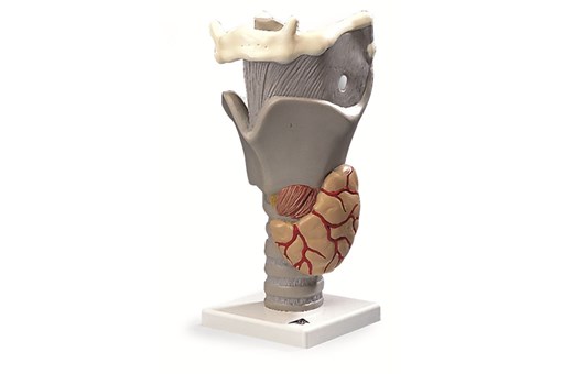
Larynx 2 Part
Specialist
Order
Order
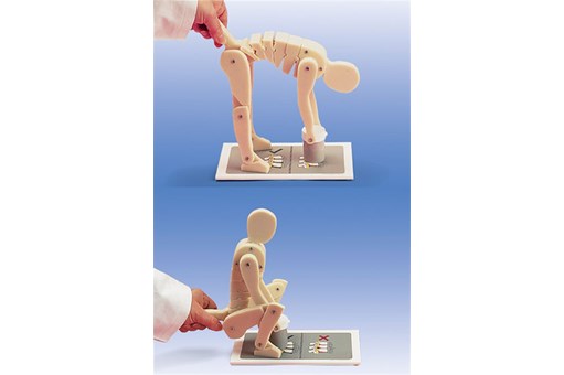
Lifting Demonstration Figure
Specialist
Order
Order
Lumbar Spinal Column Model
Specialist
Order
Order
Lung Model With Larynx 5 Part
Specialist
Order
Order
Mini Elbow Joint with Cross Section
inc gst
Mini Knee Joint with Cross Section
inc gst
Mini Shoulder Joint with Cross Section
inc gst
Mini Skeleton 'Shorty' Model
inc gst
Mr Thrifty 84cm Skeleton Model
inc gst
Multifunctional Stand
Specialist
Order
Order
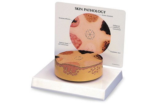
Nasco Skin Cancer Model
Specialist
Order
Order
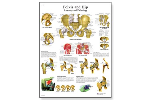
Pelvis And Hip Chart
Specialist
Order
Order
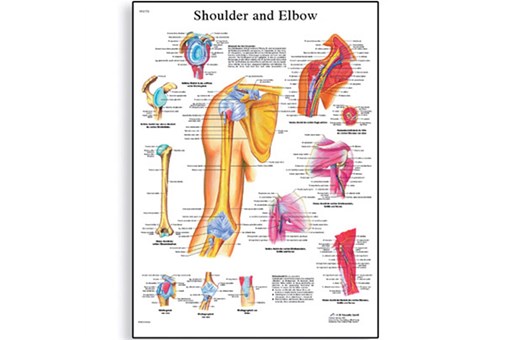
Shoulder And Elbow Chart
Specialist
Order
Order
Skin Model - 3 Part
Specialist
Order
Order
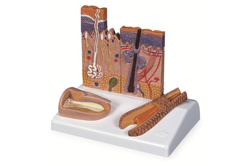
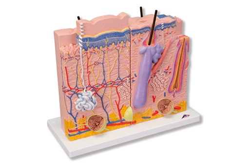
Skin Section Model
Specialist
Order
Order
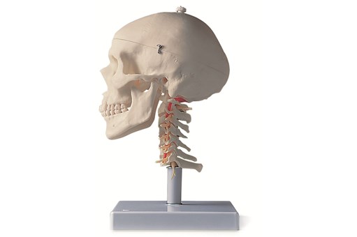
Skull Model - 4 Part
Specialist
Order
Order
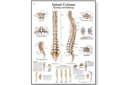
Spinal Column Chart
Specialist
Order
Order
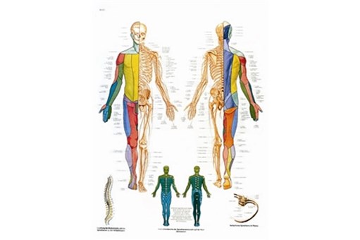
Spinal Nerves Chart
Specialist
Order
Order
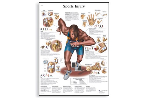
Sports Injuries Chart
Specialist
Order
Order
Stan Classical Skeleton Model On 5 ft Roller Stand
inc gst
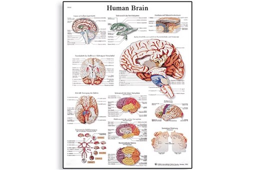
The Human Brain Chart
inc gst
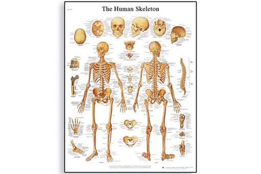
The Human Skeleton Chart
inc gst
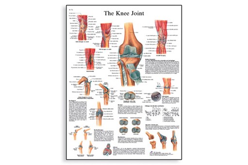
The Knee Joint Chart
Specialist
Order
Order
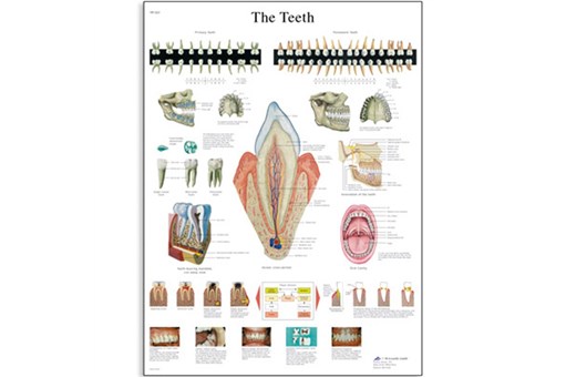
The Teeth Chart
Specialist
Order
Order
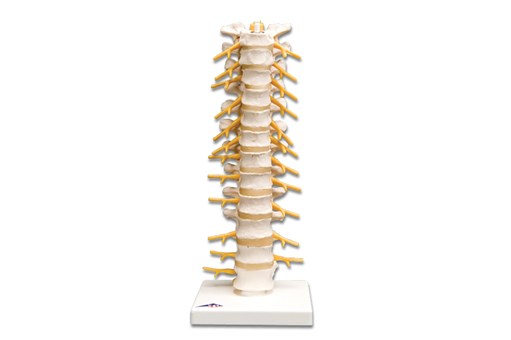
Thoracic Spinal Column Model
Specialist
Order
Order